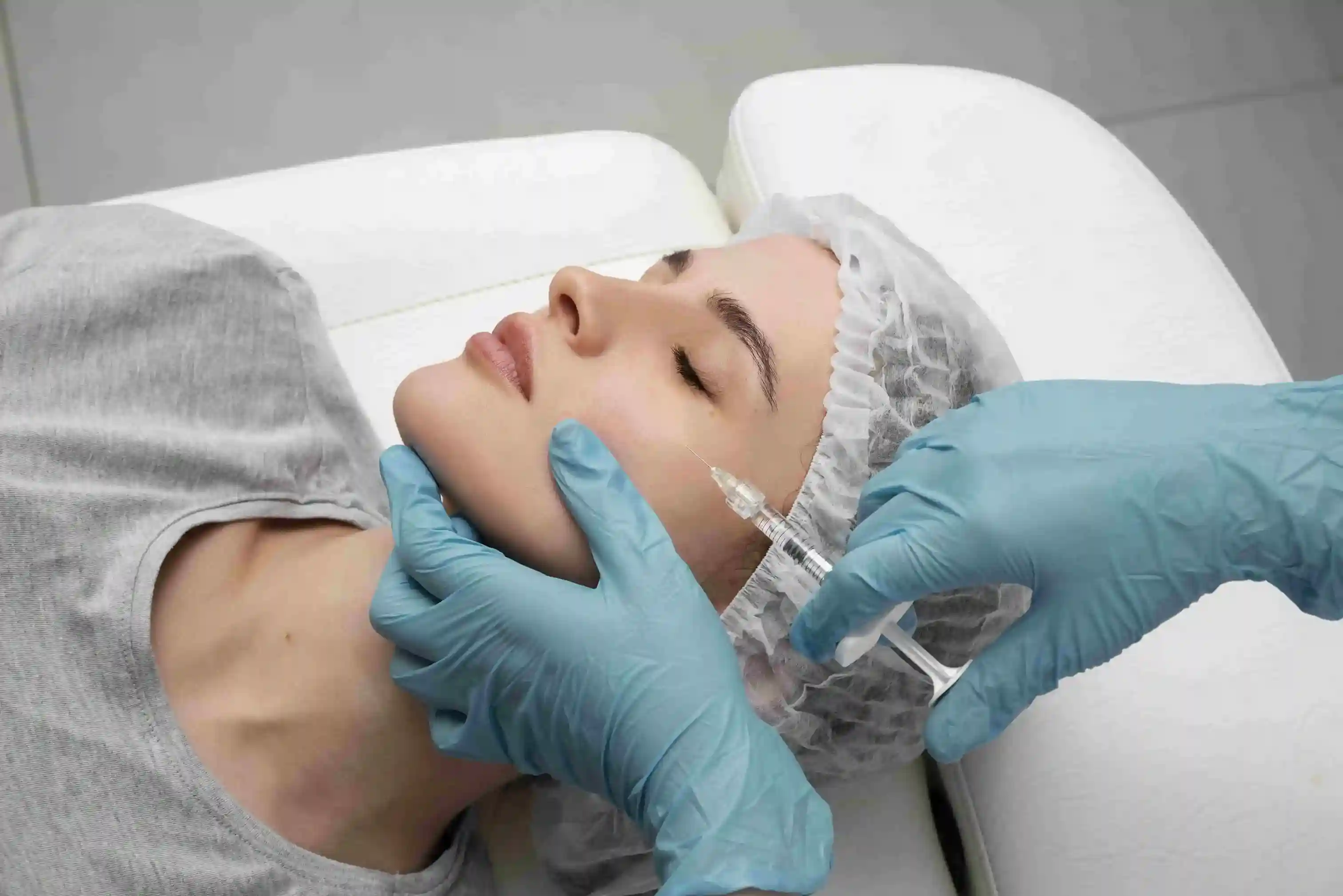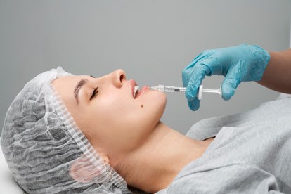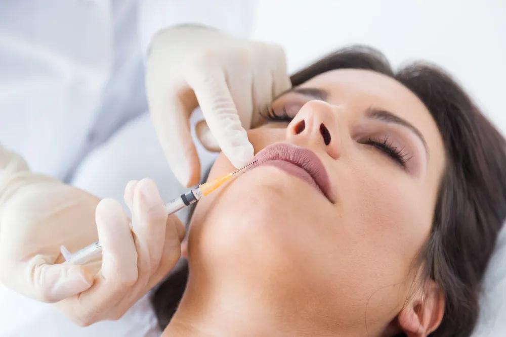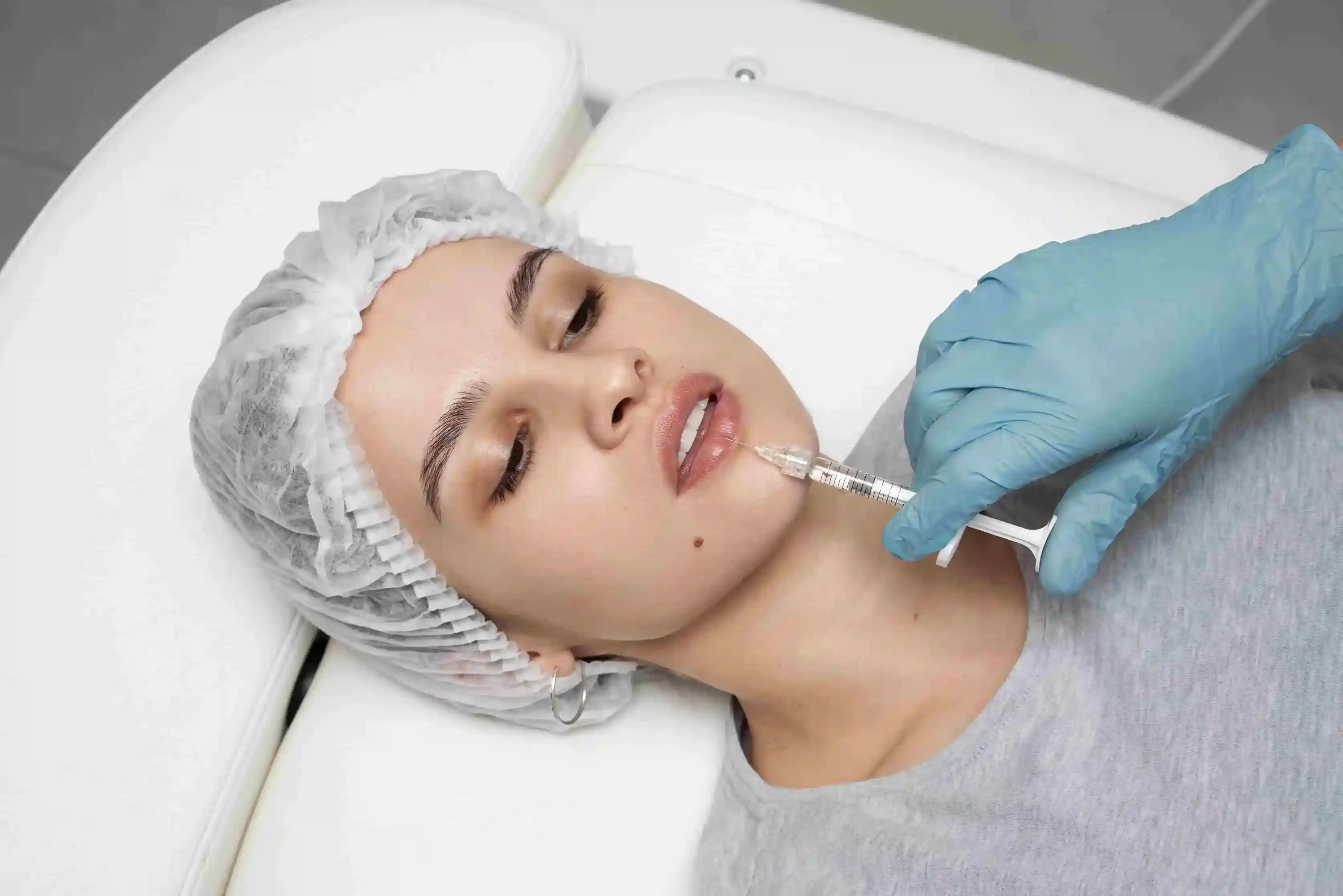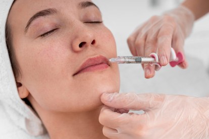
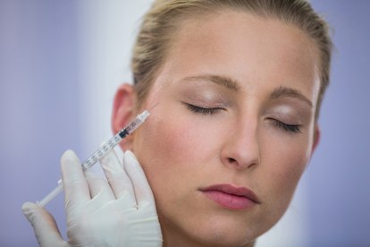
While dermal fillers are generally safe, vascular occlusion (VO) remains one of the most critical and severe complications in aesthetic medicine. According to FDA safety communications regarding dermal fillers, the unintentional injection of product into a blood vessel can lead to profound tissue ischemia and damage. Whether you are a licensed practitioner refining your emergency protocols or a patient seeking to understand post-treatment recovery, recognizing how vascular occlusion presents is an absolute necessity.
Failing to identify the early warning signs—or mistakenly brushing them off as normal lip filler swelling—can result in severe consequences, including irreversible tissue necrosis and permanent scarring. This clinical breakdown highlights the early indicators, tracks the timeline of symptom progression, and explains how to distinguish a true medical emergency from routine post-injection inflammation.
What Is Vascular Occlusion in Dermal Filler Treatments?
Vascular occlusion occurs when a dermal filler compromises local blood circulation. This happens through two primary mechanisms: intra-arterial injection (the filler directly blocks the vessel from the inside) or extrinsic compression (a large volume of filler presses against the vessel from the outside, pinching it shut).
Deprived of a vital oxygen supply, the surrounding skin and underlying structures begin to starve. While this ischemic event can occur anywhere on the face, highly vascularized areas such as the lips, nasolabial folds, and the glabella are considered primary danger zones. This risk applies to all hyaluronic acid (HA) products, whether your clinic utilizes Juvederm, Restylane, or other established brands.
Why Early Recognition Is Critical: Time is Tissue
The initial signs of vascular occlusion are notoriously subtle, often masquerading as standard injection trauma. However, unlike purple bruising after lip fillers—which typically peaks and resolves—a vascular compromise is a rapidly progressive event.
Identifying these symptoms in their infancy allows for immediate, aggressive intervention. The gold standard for reversing HA-induced occlusion is flooding the ischemic area with hyaluronidase, an enzyme that rapidly breaks down the filler, restoring perfusion and saving the tissue.
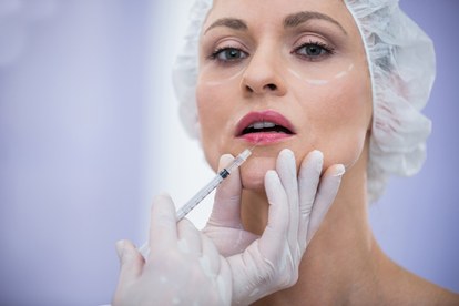
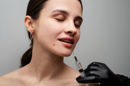
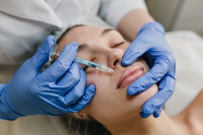
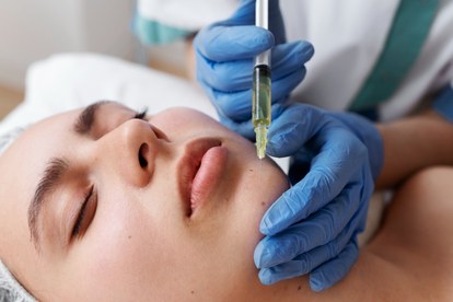
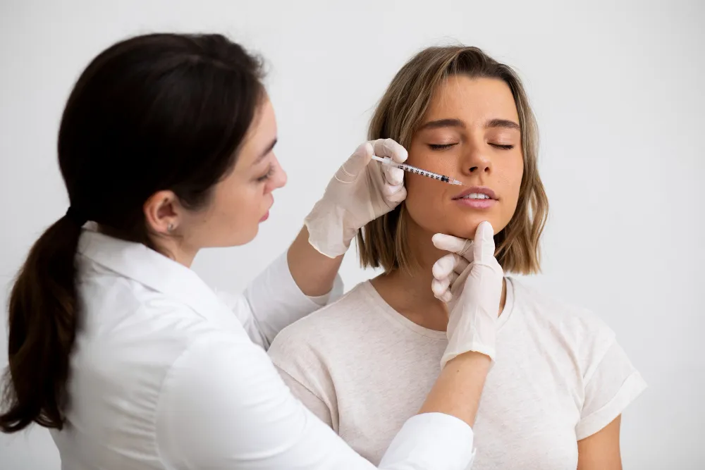
Early Signs of Vascular Occlusion in the Lips & Face
The perioral region and lips are highly reactive to fluctuations in blood supply. If an occlusion is suspected, practitioners and patients should immediately check for the following red flags:
- Disproportionate Pain: Pain that is sharp, severe, or escalating hours after the procedure, far exceeding the typical discomfort associated with standard filler placement.
- Blanching (Skin Whitening): A stark, unnatural whitening or pallor of the skin in the exact distribution area of the compromised artery.
- Prolonged Capillary Refill Time (CRT): When applying firm pressure to the skin, it naturally turns white. Healthy tissue regains its pink hue in 1-2 seconds. In ischemic tissue, this refill is significantly delayed (greater than 3 seconds) or completely absent.
- Poikilothermia (Temperature Drop): The compromised tissue will feel palpably colder to the touch when compared to adjacent, healthy skin.
Vascular Occlusion Stages & Symptom Progression
As outlined in multiple clinical studies on aesthetic complications via PubMed, untreated vascular occlusion follows a highly destructive timeline:
1. Early Ischemic Stage (0–2 hours)
This phase is marked by immediate blanching, intense localized pain, and a sluggish capillary refill. The surrounding skin may appear unusually pale or slightly mottled.
2. Progressive Stage (2–24 hours)
As hypoxia sets in, the tissue develops livedo reticularis—a dusky, bluish-purple, net-like discoloration. The area becomes increasingly indurated (firm), and the pain may intensify. Inexperienced observers frequently misdiagnose this critical stage as a severe, expanding bruise.
3. Advanced Stage / Necrosis (24+ hours)
Without enzymatic intervention, cellular death begins. Skin necrosis manifests as blackening or sloughing of the tissue, followed by ulceration. Reversing damage at this late stage is exceedingly difficult and often results in scarring.
The Phenomenon of Delayed Vascular Occlusion
While most occlusions are acute and occur while the patient is still in the chair, delayed vascular occlusion can manifest hours or even days post-treatment. This insidious onset is typically driven by:
- Progressive edema (swelling) that slowly compresses an adjacent blood vessel over time.
- The hydrophilic (water-attracting) properties of HA fillers, which draw in fluid and expand over the initial 48-hour period.
- Delayed inflammatory or immune responses that rapidly alter internal tissue pressure.
Because of this delayed risk window, providing patients with clear, written aftercare instructions and emergency contact protocols is non-negotiable.
Normal Swelling vs. Vascular Occlusion: A Diagnostic Comparison
Differentiating between a routine inflammatory response and a true vascular emergency is a vital skill in aesthetic practice.
| Diagnostic Feature | Routine Post-Filler Reaction | Vascular Emergency (Occlusion) |
|---|---|---|
| Pain Characteristics | Mild to moderate tenderness, manageable | Severe, escalating, and highly disproportionate |
| Swelling Pattern | Generally symmetrical and highly localized | Often asymmetrical, spreading along the vessel path |
| Skin Discoloration | Erythema (redness) or typical purpura (bruising) | Initial blanching, evolving into dusky, reticular netting |
| Tissue Temperature | Normal or mildly warm due to inflammation | Noticeably cool or cold to the touch |
| Capillary Refill (CRT) | Normal flush (1-2 seconds) | Severely delayed (3+ seconds) or completely absent |
| Clinical Progression | Peaks early, then gradually improves over 3-7 days | Rapidly deteriorates hour by hour |
Patient Communication and Emergency Action Protocols
Proactive communication is the strongest defense against severe complications. Patients must be educated on the exact symptoms that warrant an emergency call to their provider.
If clinical signs point to vascular compromise, practitioners should:
- Cease injecting immediately if the procedure is ongoing.
- Perform a Capillary Refill Time (CRT) assessment across the affected and adjacent areas.
- Initiate emergency protocols. Prompt assessment and aggressive high-dose flooding with hyaluronidase products are imperative. A "wait and see" approach is never acceptable when ischemia is suspected.
For more deep dives into managing aesthetic complications, explore our comprehensive FAQ section and clinical guides on Unboxed Fillers.
FAQ: Vascular Occlusion After Dermal Fillers
What is the primary mechanism behind vascular occlusion in filler treatments?
It typically occurs either through direct intra-arterial injection (the needle or cannula deposits filler inside the artery) or through extrinsic compression, where a large bolus of filler places excessive pressure on the outside of the vessel, cutting off circulation.
Can vascular occlusion present days after the initial treatment?
Yes. While acute occlusion is immediate, delayed vascular occlusion can surface 12 to 48 hours later. This is often due to the filler expanding as it attracts water, or secondary swelling eventually compressing a nearby vessel.
What does a lip vascular occlusion look like early on?
The hallmark early sign is a stark white patch (blanching) immediately post-injection. If untreated, this evolves into a dusky, grayish-blue, or purplish mottled appearance, usually accompanied by intense, throbbing pain that extends beyond the lips.
What is the standard reversal treatment for occlusion?
The universal emergency protocol involves injecting high doses of hyaluronidase directly into and around the ischemic area. This enzyme quickly dissolves the hyaluronic acid filler, relieving the blockage and restoring vital blood flow.
When should a patient seek immediate medical intervention?
Contact your aesthetic provider or seek emergency care immediately if you experience sharp, escalating pain, notice your skin turning pale white or grayish-blue, feel unnatural coldness in the injected area, or if symptoms aggressively worsen over a few hours.
Professional Disclaimer: This content is provided for educational and informational purposes only. It is intended for licensed medical professionals and informed readers. It does not provide treatment protocols, emergency management instructions, or clinical dosing guidance. Please review our full Disclaimer and Terms of Use.